The Ultimate Guide to E. Coli: 5 Microscope Tips

Escherichia coli, commonly known as E. coli, is a bacterium that often gets a bad reputation due to its association with foodborne illnesses. However, it's essential to understand that not all E. coli strains are harmful; in fact, these bacteria play crucial roles in various ecosystems and even our digestive systems. This comprehensive guide aims to delve into the world of E. coli, offering expert insights and practical tips for observing and studying this fascinating microorganism under the microscope.
Unveiling the World of E. Coli

E. coli belongs to the family Enterobacteriaceae, a diverse group of Gram-negative bacteria that inhabit various environments, from soil to the intestinal tracts of animals. While some E. coli strains are harmless and even beneficial, others can cause severe illnesses. This diversity makes studying E. coli an intriguing and essential task for microbiologists and public health professionals alike.
The microscopic world of E. coli offers a captivating glimpse into the complexity of bacterial life. With the right techniques and tools, scientists can unravel the secrets of these tiny organisms, leading to breakthroughs in medicine, agriculture, and environmental science. This guide aims to equip you with the knowledge and skills to observe and analyze E. coli effectively, contributing to a deeper understanding of this ubiquitous bacterium.
Microscope Preparation and Sample Collection

To embark on your journey of exploring E. coli, proper microscope preparation and sample collection are crucial. Here’s a step-by-step guide to ensure you’re ready for your microscopic adventure:
Choosing the Right Microscope
Selecting an appropriate microscope is the first step. For observing E. coli, a compound microscope with a magnification range of 40x to 100x is ideal. Ensure your microscope is equipped with a bright light source, as E. coli is relatively small and requires adequate illumination for clear visualization.
Sample Collection and Preparation
E. coli can be found in various environments, including soil, water, and the intestinal tracts of animals. For laboratory studies, you can collect samples from these sources or use commercially available E. coli cultures. Ensure you follow proper safety protocols when handling potential pathogens, such as wearing gloves and working in a sterile environment.
Once you have your sample, prepare a bacterial smear slide. This involves spreading a small amount of the sample onto a glass slide, allowing it to air dry, and then gently heating it over a flame to fix the bacteria to the slide. This process ensures the bacteria remain in place during staining and observation.
Staining Techniques
Staining is an essential step in visualizing E. coli under the microscope. Gram staining, a widely used technique for bacterial differentiation, is particularly effective for E. coli. Here’s a simplified guide to Gram staining:
- Primary Stain: Apply a drop of crystal violet solution to the slide and allow it to stain the bacteria for about 1 minute. Crystal violet will penetrate the cell walls of Gram-positive bacteria, turning them purple.
- Mordant: Add a drop of Gram's iodine solution to the slide. Iodine acts as a mordant, enhancing the penetration of the primary stain.
- Decolorization: Quickly rinse the slide with alcohol to remove the excess stain. This step is crucial, as it washes away the primary stain from Gram-negative bacteria, including E. coli.
- Counterstain: Apply a drop of safranin or carbol fuchsin solution to the slide. These counterstains color the Gram-negative bacteria, making them visible under the microscope.
- Observation: Rinse the slide with water and observe under the microscope. E. coli, being a Gram-negative bacterium, will appear pink or red against a contrasting background.
Observing E. Coli: Tips and Tricks
Now that your microscope is prepared and your samples are ready, it’s time to dive into the world of E. coli. Here are some expert tips to enhance your observation experience:
Focus and Magnification
Start with a low magnification (40x) to get a general idea of the sample’s distribution. Then, gradually increase the magnification to 100x for a more detailed view. Ensure your microscope is properly focused at each magnification to obtain clear images of the bacteria.
Cell Morphology
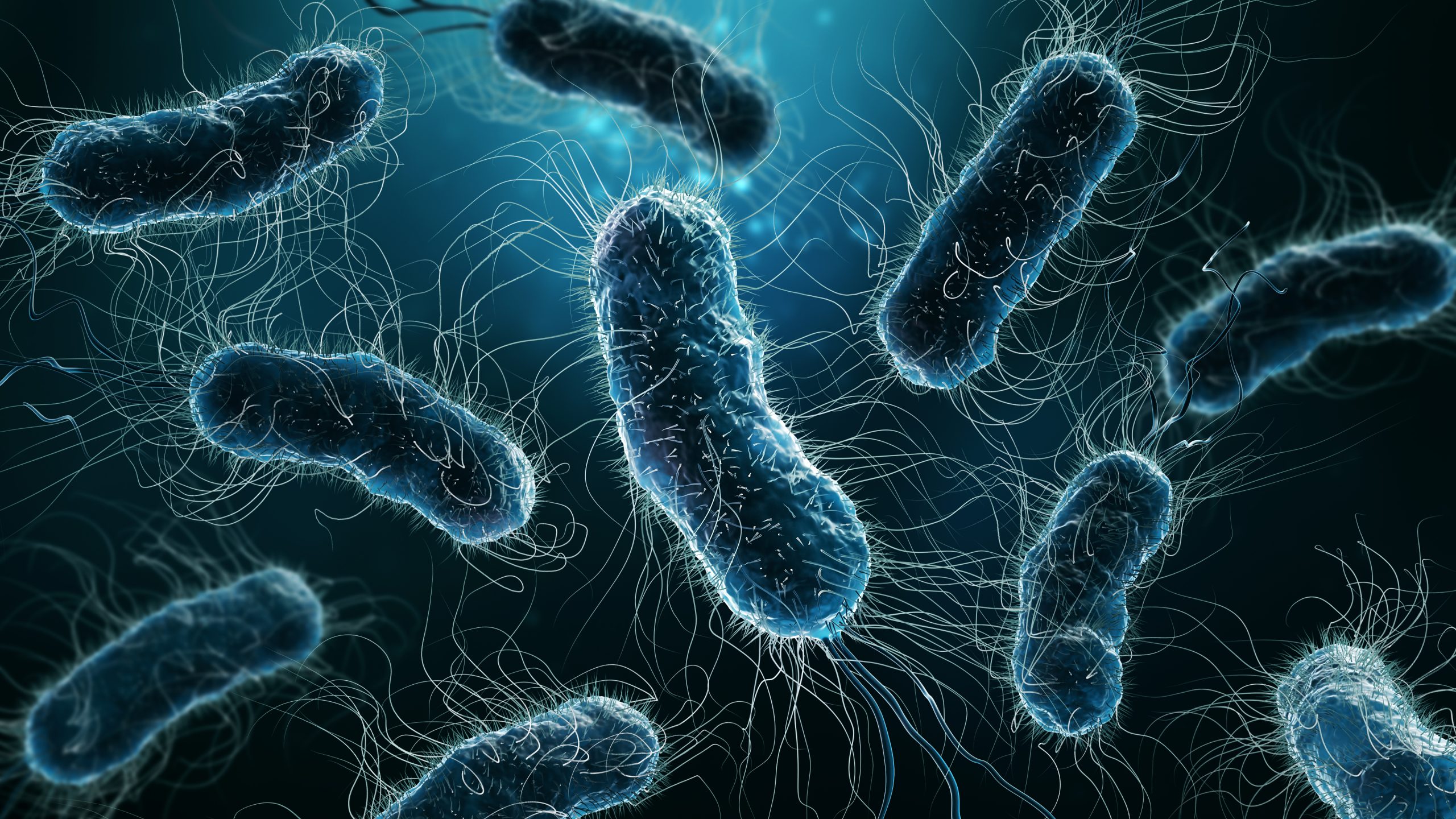
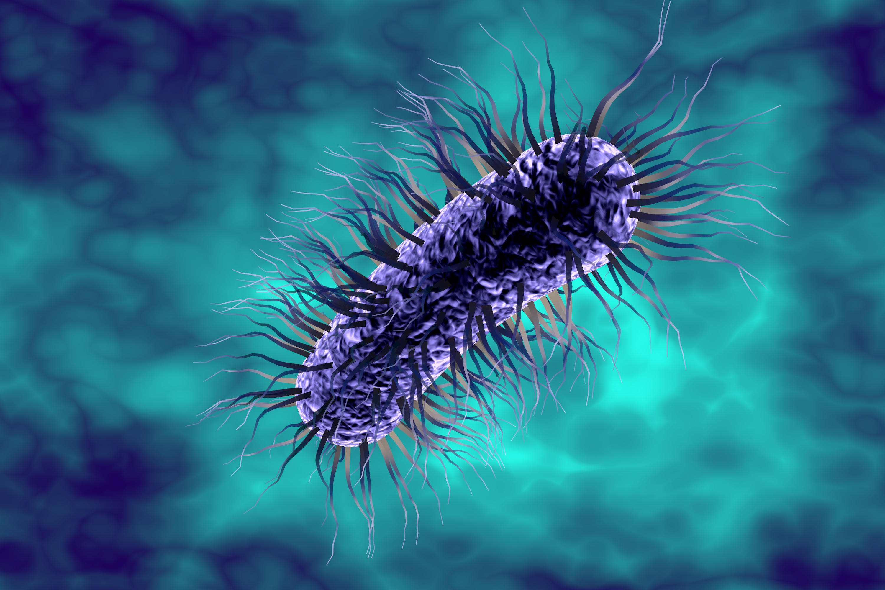
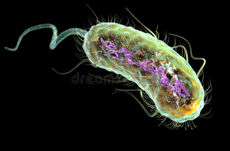
E. coli typically exhibits a rod-shaped morphology, often appearing as short, straight rods. However, under certain conditions, they can also adopt a coccoid form. Pay attention to the size, shape, and arrangement of the bacteria. This morphological analysis can provide insights into the health and behavior of the E. coli population.
Colony Characteristics
If you’re working with cultured E. coli, observe the characteristics of the bacterial colonies. E. coli colonies often appear as circular, smooth, and convex with a shiny surface. The color and texture of the colonies can vary depending on the strain and growth conditions. Documenting these colony characteristics can be valuable for strain identification and comparison.
Motility and Movement
E. coli is known for its motility, thanks to its flagella. Under the microscope, you might observe the bacteria moving in a characteristic darting or tumbling motion. This motility is a fascinating feature to observe and can provide insights into the behavior and response of E. coli to its environment.
Comparison with Other Bacteria
To enhance your understanding of E. coli, consider observing other bacterial species alongside it. This comparative analysis can help you appreciate the unique characteristics of E. coli and develop a broader understanding of bacterial diversity. For example, compare E. coli with a Gram-positive bacterium like Staphylococcus aureus to appreciate the differences in morphology and staining behavior.
Practical Applications and Future Implications
Studying E. coli under the microscope is not just an academic exercise; it has practical applications and far-reaching implications. Here’s a glimpse into the world of E. coli beyond the microscope:
Food Safety and Public Health
E. coli is often associated with foodborne illnesses, making its study crucial for food safety and public health. By understanding the behavior and characteristics of different E. coli strains, scientists can develop better detection methods and prevention strategies. This knowledge is vital for ensuring the safety of our food supply and protecting public health.
Environmental Impact
E. coli plays a significant role in environmental processes, particularly in nutrient cycling and decomposition. By observing and studying E. coli in various ecosystems, researchers can gain insights into the health of our environment. This knowledge can inform conservation efforts and sustainable practices, ensuring the long-term health of our planet.
Biotechnology and Medicine
E. coli has been a workhorse in biotechnology and medical research for decades. Its ability to grow rapidly, its well-characterized genome, and its ease of manipulation make it an ideal model organism. By studying E. coli under the microscope, scientists can gain insights into fundamental biological processes, leading to advancements in genetic engineering, drug development, and biomedical research.
| E. coli Strain | Application |
|---|---|
| K-12 | Biotechnology, Genetic Engineering |
| O157:H7 | Food Safety, Public Health |
| Nissle 1917 | Probiotic, Gut Health |

Conclusion: A Microscopic Adventure Awaits

Observing E. coli under the microscope is a captivating journey into the microscopic world. With the right techniques and a curious mind, you can unlock the secrets of this ubiquitous bacterium. This guide has equipped you with the knowledge and skills to embark on your own microscopic adventure, contributing to a deeper understanding of E. coli and its impact on our world.
Frequently Asked Questions
How do I ensure proper sample collection for E. coli observation?
+To collect samples for E. coli observation, follow these steps: Wear gloves and collect samples from potential sources like soil, water, or fecal matter. Ensure you work in a sterile environment to prevent contamination. If collecting from water, use a sterile container and filter the sample. If working with fecal matter, use a sterile swab. Once collected, prepare a bacterial smear slide for observation.
What safety precautions should I take when handling E. coli samples?
+When handling E. coli samples, especially those from potential sources of infection, follow these safety precautions: Wear personal protective equipment (PPE) like gloves, lab coats, and eye protection. Work in a biosafety cabinet or a designated sterile area. Avoid touching your face or mouth while handling samples. Dispose of used materials properly, following institutional guidelines for biohazardous waste.
Can I observe E. coli without staining?
+While staining enhances the visibility of E. coli under the microscope, it is possible to observe unstained samples. However, the contrast between the bacteria and the background may be lower, making visualization more challenging. Staining is recommended for clearer and more accurate observations.
How do I identify different E. coli strains under the microscope?
+Identifying different E. coli strains under the microscope can be challenging due to their similar morphology. However, you can observe certain characteristics such as colony morphology, motility patterns, and growth rate. Additionally, molecular techniques like PCR and DNA sequencing can provide more definitive strain identification.


